Technology

Our Technology

Cutting-Edge Technology for Predictable Endodontic Success

The difference between conventional root canal treatment and specialist endodontic care lies fundamentally in visualization and precision. At our Leicester practice, we leverage state-of-the-art technology to overcome anatomical challenges and deliver minimally invasive, predictable outcomes.

The Endodontic Operating Microscope

Microscope-Assisted Endodontics: The Specialist Standard

Every procedure is performed under a high-magnification dental operating microscope. This isn't merely an accessory it's an essential tool that transforms endodontics from a procedure performed predominantly by feel into one driven by direct visual control.

The Microscope Advantage:

Enhanced Visualization: High-resolution magnification provides over 900% greater visual clarity than the unaided eye, allowing precise identification of:

  • Tiny or calcified canals frequently missed in conventional treatment
  • Micro-fractures and structural defects
  • Anatomical variations requiring specialized navigation
  • Separated instruments requiring delicate retrieval

Precision and Predictability:

  • Enables minimally invasive procedures
  • Reduces risk of procedural errors
  • Minimizes damage to surrounding tissue
  • Facilitates faster, more comfortable recovery
  • Critical for complex retreatment cases

What This Means for You:

The microscope allows us to see what others cannot, enabling successful treatment of cases deemed "too difficult" by conventional standards. Whether locating a fourth canal in a molar, retrieving a separated instrument, or diagnosing a hairline fracture, the microscope provides the visual information necessary for confident, accurate intervention.

Dynamic 3D Navigation (NaviDent)

NaviDent: The Future of Complex Endodontics

For cases involving extremely difficult anatomy severe calcification, deep angulation, complex root configurations Dr. Nathani utilizes Dynamic 3D Navigation technology, specifically the NaviDent system. This represents the most advanced dynamic navigational system available in dentistry today.

How Dynamic Navigation Works:

The NaviDent system integrates a pre-operative Cone Beam CT (CBCT) scan with real-time tracking software, allowing digital surgical planning with sub-millimeter accuracy. During treatment, the system continuously tracks the position, angle, and depth of the dental instrument, providing real-time guidance to the exact target area.

Revolutionary Benefits:

Minimally Invasive Access:

  • Achieves precise access cavity preparation
  • Conserves maximum healthy tooth structure
  • Enables accurate treatment of severely calcified canals

Enhanced Confidence in Complexity:

  • Provides certainty and control for highly complex cases (Complexity Codes 2 and 3)
  • Successfully treats cases with twisted roots, severe angulation, and calcification
  • Offers tangible assurance for patients who've experienced previous treatment failure

Who Benefits from NaviDent Navigation?

  • Failed previous root canal due to missed canals
  • Severely calcified root canals
  • Complex anatomical variations
  • Retreatment cases with difficult access
  • Patients seeking the highest precision available

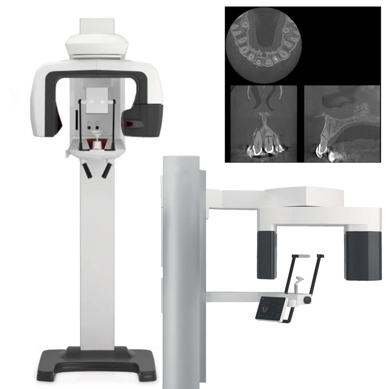
3D CT Scan (J Morita CBCT)

J Morita CBCT: High Definition 3D Diagnosis in Leicester

For complex endodontic cases, precise diagnosis is everything. At our Leicester practice, Dr. Nathani uses a state of the art J Morita Cone Beam CT (CBCT) scanner to capture highly detailed 3D images of teeth, roots, and the surrounding bone. Many practices use standard scanners, but J Morita imaging is widely regarded as among the best in dentistry, giving exceptional clarity for accurate diagnosis and careful treatment planning.

The 3D Scan Advantage:

True 3D Visualisation: A detailed 3D view helps reveal critical findings, including:

  • Hidden or extra canals that can be missed on 2D imaging
  • Root fractures and micro cracks
  • Root resorption, internal or external
  • The exact size and location of infection around the root tip
  • Unusual root anatomy, curvature, or complex canal systems

Precision Planning and Safety:

  • More accurate treatment planning for complex cases
  • Reduced risk of surprises during treatment
  • Better decision making for retreatment and surgical cases
  • Clearer communication, you can see what we see

What This Means for You:

Having advanced J Morita 3D scanning available here in Leicester means you can benefit from a higher level of diagnostic detail without being referred elsewhere. Whether you have persistent symptoms, a previously failed root canal, suspected fracture, or unusual anatomy, 3D imaging helps us identify the true cause, choose the safest approach, and improve the chance of long term success.